Understanding biomagnetic beads in one article
Magnetic beads can separate biomolecules easily and effectively. Use this guide to compare different surface chemical modifications and find the type that suits your application.
What are magnetic beads?
Magnetic beads are composed of tiny iron oxide particles (20 to 30 nm), such as magnetite (Fe3O4), which are superparamagnetic. Superparamagnetic beads are different from common ferromagnets because they only exhibit magnetism in the presence of an external magnetic field. This property depends on the size of the particles in the beads, and allows the beads and any material they bind to be suspended and separated. Since they do not attract each other outside the magnetic field, they can be used without worrying about unnecessary clumping.
There are many types of magnetic beads available. Different surface coatings and chemical properties give each type of bead its own binding properties, which can be used for the magnetic separation (separation and purification) of nucleic acids, proteins or other biomolecules in an easy, effective and scalable manner.
This ease of use makes it easy to automate and is ideal for a range of applications, including sample preparation for next-generation sequencing (NGS) and PCR, protein purification, molecular and immunodiagnosis, and even magnetic activated cell sorting (MACS) Wait.
What is magnetic separation?
Magnetic separation uses a magnetic field to separate micron-sized paramagnetic particles from a suspension. In molecular biology, magnetic beads provide a simple and reliable method to purify various types of biomolecules, including genomic DNA, plasmids, mitochondrial DNA, RNA and proteins. The main advantage of using magnetic beads is that you can directly separate nucleic acids and other biomolecules from crude samples and various types of samples.

How does magnetic bead DNA extraction work?
Magnetic beads have existed for decades. As evidenced by the 1990 US patent, their potential in nucleic acid purification has been recognized.
After binding the DNA, the external magnetic field attracts the magnetic beads to the outer edge of the tube, thereby fixing it. When the beads are fixed, the DNA bound to the beads is retained during the washing step. The elution buffer is added and the magnetic field is removed, and then the DNA is released to become a purified sample, ready for quantification and analysis.
This method eliminates the need for vacuum or centrifugation. This method minimizes the shearing force on the target molecule, requires fewer steps and reagents than other DNA extraction schemes, and is suitable for 24, 96 and 384-well plates. automation.
Therefore, it is no wonder that magnetic beads are becoming more and more popular. In fact, manufacturers have now developed many commercial nucleic acid separation kits based on magnetic beads. They have a variety of surface chemistry and multiple application options.
Comparison of surface chemistry and application of magnetic beads
Carboxylic acid modified magnetic beads
Combination method
(1) It can be directly captured by combining with nucleic acid.
(2) Surfaces suitable for covalent bonding.
(3) It can capture molecules containing amino groups.
Application field
(1) Covalent connection
(2) Affinity purification
(3) Nucleic acid separation and purification
(4) NGS

Anti-amine magnetic beads
Combination method
(1) Surfaces suitable for covalent bonding.
(2) Non-surfactant, non-protein blocking surface.
(3) Low non-specific binding.
Application field
Conjugation application, similar to carboxylate modified beads.
Oligo (dT) coated magnetic beads
Combination method
(1) Hybridize with mRNA poly-A tail.
(2) High colloid stability.
Application field
(1) mRNA extraction and purification
(2) Reverse transcription PCR
(3) cDNA library construction
(4) NGS (RNA sequencing)
Streptavidin-coated magnetic beads
Combination method
(1) Binding biotinylated ligands, such as proteins, nucleic acids and peptides.
(2) Covalently bound streptavidin coating.
(3) Fast reaction kinetics.
(4) Low non-specific binding.
(5) High throughput and precision.
Application field
For sample preparation and assay development for genomics and proteomics.
Streptavidin-blocked magnetic beads
Combination method
(1) Binding biotinylated ligands, such as proteins, nucleic acids and peptides.
(2) Non-surfactant, non-protein blocking surface.
(3) Compared with non-streptavidin-coated beads, non-specific binding is lower by additional blocking of non-specific binding sites.
Application field
(1) Molecular and immunological diagnosis
(2) NGS library preparation
NeutrAvidin™ coated magnetic beads
Combination method
(1) Binding biotinylated ligands, such as proteins, nucleic acids and peptides.
(2) Fast reaction kinetics.
(3) Low non-specific binding.
(4) High throughput and precision.
Application field
For sample preparation and assay development for genomics and proteomics.
Protein A/G Magnetic Beads
Combination method
(1) Combine IgA and IgG protei
(2) Coating based on IgA/IgG fusion protein.
(3) Extensive binding function.
Application field
(1) Affinity purification and pull-down
(2) Immunoprecipitation
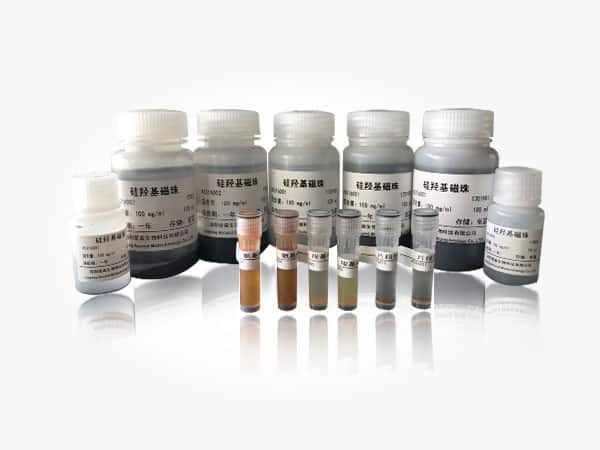
Silica coated magnetic beads
Combination method
(1) Reversibly bind nucleic acid based on salt concentration.
(2) Monodisperse particles with a size range of 400 µm or 700 µm.
Application field
Nucleic acid extraction for molecular diagnostic applications (eg qPCR).
Sepharose magnetic beads
Combination method
(1) Wide selection of ligands.
(2) Porous, providing larger surface area than other magnetic beads.
Application field
Affinity purification or capture
Immunoprecipitation


